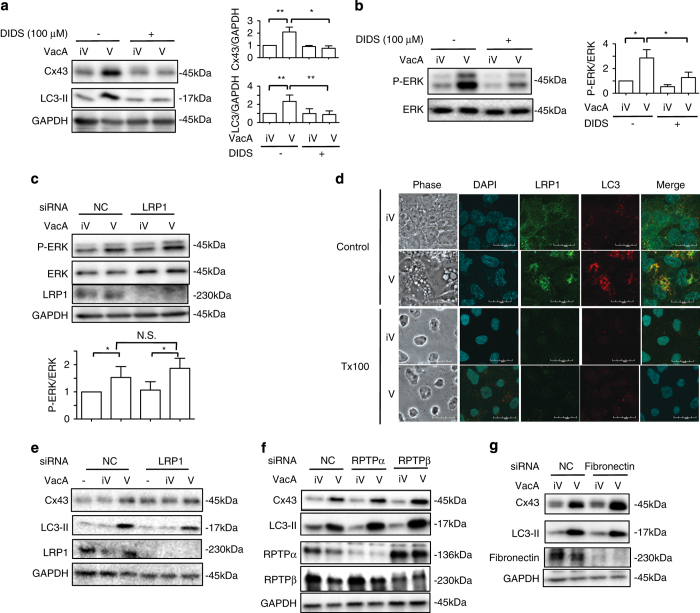
Figure 6.
VacA channel activity, not LRP1, regulates VacA-induced Cx43 and LC3-II generation via ERK activation. (a) AZ-521 cells were pretreated with or without 100 μM DIDS for 30 min and then incubated with 120 nM heat-inactivated (iV) or wild-type VacA (V) for 10 h. Cells were lysed with 1× SDS sample buffer and analyzed by immunoblotting with the indicated antibodies. Quantification of VacA-induced Cx43 and LC3-II generation in AZ-521 cells was performed by densitometry (right panel). Data are presented as mean±S.D. of values from three experiments and significance is *P<0.01, **P<0.05. Three independent experiments were performed with similar results. (b) AZ-521 cells were pretreated with control (DMSO) or 100 μM DIDS and then 120 nM heat-inactivated (iV) or wild-type VacA (V) for 1 h at 37 °C. Cells were lysed with 1× SDS sample buffer for immunoblotting with p-ERK and ERK as a loading control. Quantification of p-ERK in AZ-521 cells was performed by densitometry (right panel). Data are presented as mean±S.D. of values from three experiments and significance is *P<0.05. Experiments were repeated independent three times. (c) Control (NC) or LRP1 siRNA-transfected cells were incubated with 120 nM heat-inactivated (iV) or wild-type VacA (V) for 1 h at 37 °C. Cells were lysed with 1× SDS sample buffer for immunoblotting with anti-ERK, anti-p-ERK and anti-LRP1 antibodies. GAPDH served as a loading control. Experiments were repeated three times with similar results. (d) AZ-521 cells were incubated with 120 nM heat-inactivated (iV) or wild-type VacA (V) for 10 h and were treated with or without cold 1% T×100, fixed with 4% PFA, and reacted with anti-LRP1 (green), anti-LC3 antibodies (red) and incubated with DAPI (cyan) as described in Materials and Methods. Bars represent 20 μm. Experiments were repeated three times with similar results. (e–g) The indicated siRNA-transfected cells were incubated with 120 nM heat-inactivated (iV) or wild-type VacA (V) for 10 h at 37 °C. Cells were lysed with 1× SDS sample buffer and analyzed by immunoblotting with the indicated antibodies. GAPDH served as a loading control. Experiments were repeated three times with similar results.

