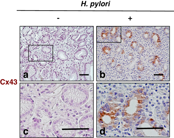
Figure 7.

H. pylori infection is associated with increased Cx43 expression in human gut tissues. Cx43 was detected (i.e., brown staining) in H. pylori-negative (a and c) and -positive (b and d) gastric mucosa. Blue color indicated nuclear staining by hematoxylin. The black square shows low magnification views of the structures in c and d, respectively (a and b: ×20 ; c and d: ×80). The figure shows one experimental result representative of 5 H. pylori-negative and 11 of H. pylori-positive samples. Statistically significant difference between H. pylori-negative and -positive mucosa was observed (P=0.0256). Fisher’s exact test, H. pylori-positive versus -negative mucosa. Bars represent 50 μm.
