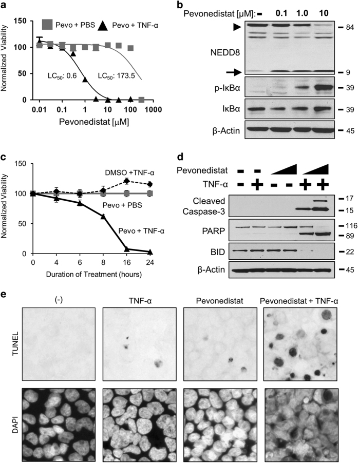
Figure 1.
Pevonedistat+TNF-α is cytotoxic. (a) Cultured rat H-4-II-E cells were treated with pevonedistat in combination with either PBS (gray boxes) or 5 ng/ml TNF-α (black triangles) for 24 h. The LC50 was determined using the least-squares method, and solid lines indicate a non-linear fit of the data. (b) Cells were treated with DMSO (−) or pevonedistat (0.1, 1.0, and 10 μM) for 8 h. Lysates were western blotted for the indicated proteins. NEDD8-cullin (arrowhead) and unbound NEDD8 (arrow) are indicated (c). Viabilities were determined at the indicated time points after treatment with DMSO+5 ng/ml TNF-α (black circles), 10 μM pevonedistat+PBS (gray boxes), or 10 μM pevonedistat+5 ng/ml TNF-α (black triangles). (d) Lysates of cells treated with 1 or 10 μM of pevonedistat±TNF-α for 8 h were western blotted for the indicated apoptotic marker proteins. (e) TUNEL (terminal deoxinucleotidyl transferase-mediated dUTP-fluorescein nick end labeling; upper) and DAPI (4,6-diamidino-2-phenylindole; lower) staining of apoptotic cells after 6 h of treatment. All viability experiments were performed in triplicate and error bars indicate±S.E.M. Approximate molecular sizes of proteins (in kDa) are given to the right of blots.

