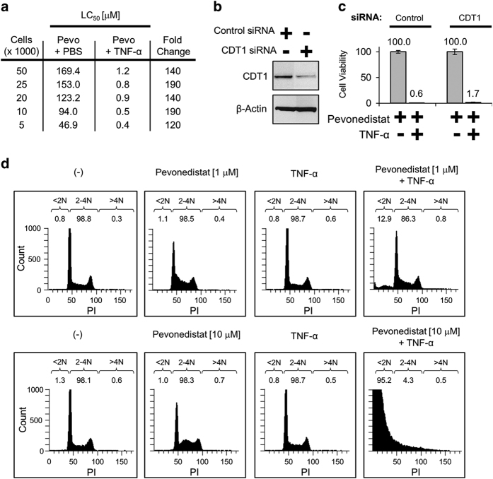
Figure 2.
DNA re-replication does not drive pevonedistat+TNF-α toxicity. (a) H-4-II-E cells were seeded from sparse (5000) to confluent (50 000) in wells of a 96-well plate. Cells were treated with pevonedistat±TNF-α, and viabilities were determined after 24 h. (b) Cells were transfected with either a siRNA pool against a non-targeting control or a single oligonucleotide siRNA against CDT1. Lysates were collected 5 days later and western blotted for the indicated proteins. (c) Viabilities of cells transfected with control or CDT1 siRNAs treated with pevonedistat±TNF-α were determined after 48 h. Viability experiments were performed in triplicate and error bars indicate±SEM. (d) Actively dividing cells were treated with 1 μM pevonedistat (upper) or 10 μM pevonedistat (lower)±TNF-α for 8 h. Cells were then pulsed with Brd-U and fixed, and DNA content was determined via FACS analysis. Displayed are cells that stained positive for PI versus the count of Brd-U positive. DNA content was determined by sorting cells into <2N, 2–4N, and >4N groups and displayed as a percentage of the total live cells counted.

