Figure 3.

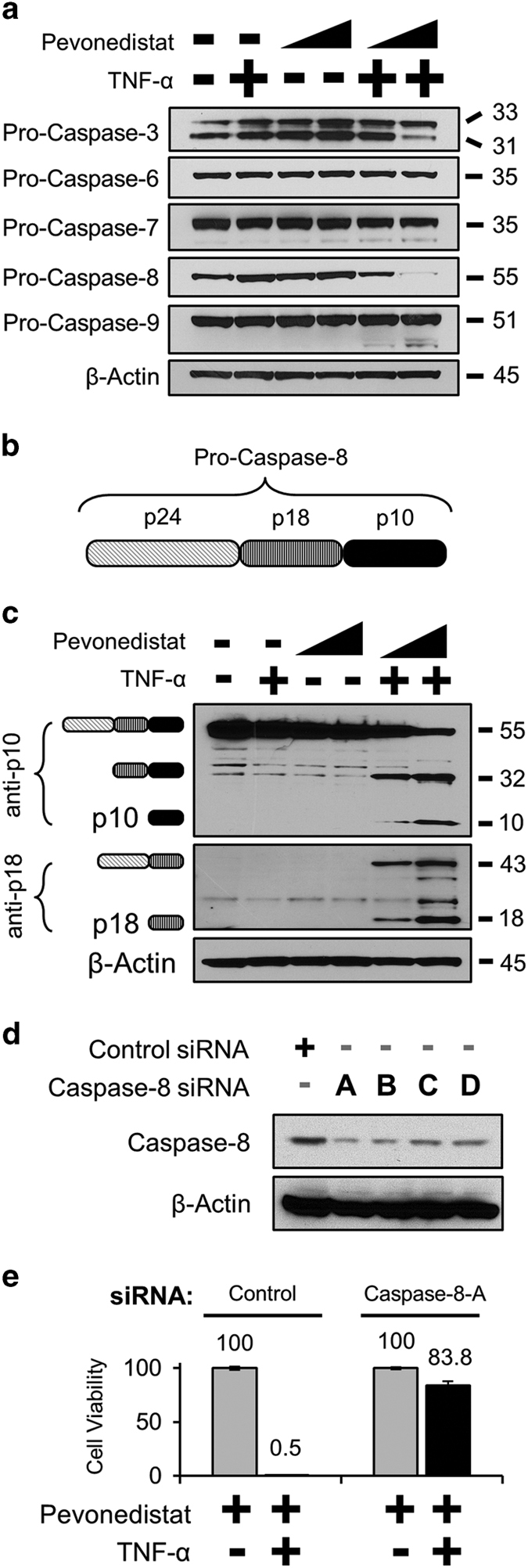
Pevonedistat+TNF-α cytotoxicity is mediated by caspase-8. (a) H-4-II-E cells were treated with 1 or 10 μM pevonedistat±TNF-α for 16 h. Extracts were western blotted for the pro-enzyme form of the indicated caspases. (b) The schematic of the individual subunits of pro-caspase-8 (p24, p18, and p10) are as indicated. (c) Lysates from cells treated with 1 or 10 μM pevonedistat±TNF-for 8 h were western blotted with antibodies specific for epitopes within the caspase-8 p10 (top) or p18 subunits. The predicted caspase-8 subunits are indicated to the left of the image based on the expected size of the product. (d) Lysates from cells transfected with siRNA oligonucleotides against either a non-targeting control or against caspase-8 were western blotted for full-length caspase-8. (e) Cells were transfected with either a non-targeting control or the caspase-8-A siRNA. Four days later, cells received the indicated treatments and viability was assessed after an additional 48 h. All viability experiments were performed in triplicate, and error bars indicate±S.E.M. Approximate molecular sizes of proteins (in kDa) are given to the right of blots.
