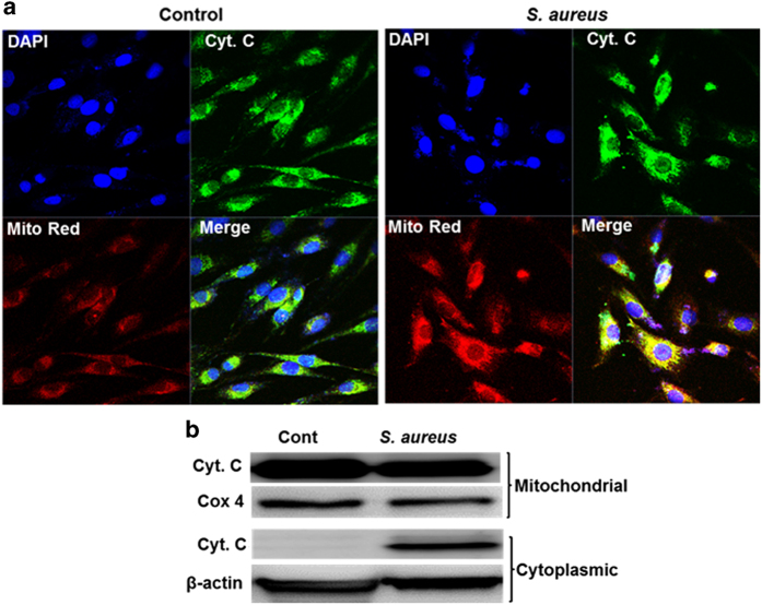
Figure 3.
S. aureus-challenged retinal Müller glia releases cytochrome c from mitochondria. MIO-M1 cells were left uninfected (control) or challenged with S. aureus (SA) RN6390 (multiplicity of infection, 10 : 1) for 8 h. Cells were stained with MitoTracker red (Mito Red) dye followed by immunostaining for cytochrome c and observed under confocal microscope (a). In another experiment, S. aureus-challenged MIO-M1 cells were subjected to subcellular fractionation followed by western blot analysis of cytochrome c (b). Cox 4 and β-actin antibodies were used as protein loading controls for mitochondrial and cytoplasmic fractions, respectively. Results are representative of two independent experiments.

