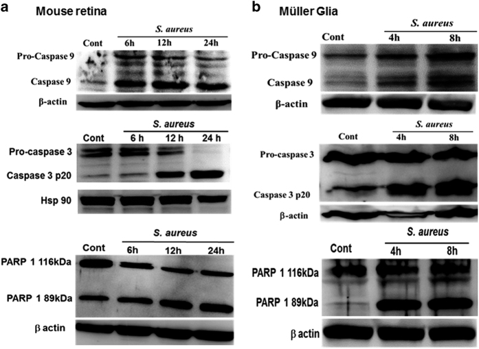
Figure 5.
S. aureus infection initiates the activation of caspase-9 and -3 and the cleavage of PARP-1 in the mouse retina and retinal Müller glia. The retinal lysates, prepared from S. aureus-infected B6 mouse eyes at the indicated time point post infection were subjected to western blot analysis using caspase-9, caspase-3 and PARP-1-specific antibodies. The retinal tissue from PBS-injected eyes at 24 h was used as control (a). For in vitro studies, MIO-M1 cells were left uninfected (control) or challenged with S. aureus RN6390 (multiplicity of infection, 10 : 1) for indicated time periods. Cell lysates were prepared using radioimmunoprecipitation assay buffer containing protease and phosphatase inhibitors cocktail was used for the detection of caspase-9 and caspase-3 by western blot (b). Results are representative of two independent experiments.

