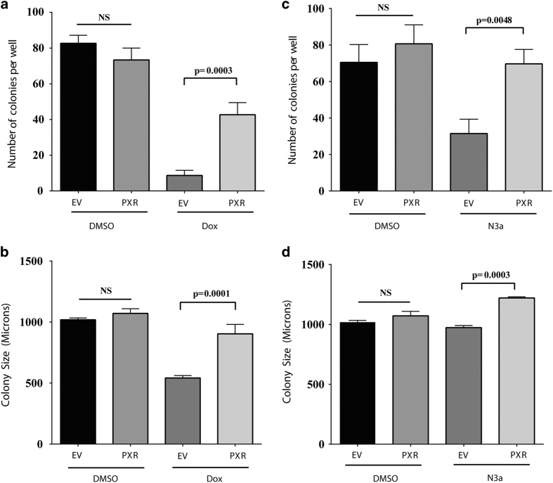
Figure 4.
PXR expression protects against doxorubicin and nutlin-3a toxicity contributing to malignant transformation. (a) HCT116 (p53+/+) cells stably transduced with lentiviral FLAG-tagged EV or FLAG-tagged PXR (PXR) were seeded at 5×10⁴ cells/well and incubated for 10 days with doxorubicin (100 nM) (Dox). The resulting colonies were counted using light microscopy (×4 magnification). (b) HCT116 (p53+/+) cells stably expressing EV or PXR were incubated for 10 days with doxorubicin (100 nM) (Dox). The size of colony foci (in micrometers) was measured using the ROI perimeter tool in the cellSens software (Olympus). (c) HCT116 (p53+/+) cells stably transduced with EV or PXR were seeded at 5×10⁴ cells/well and incubated for 10 days with nutlin-3a (1 μM) (N3a). The resulting colonies were counted using light microscopy (×4 magnification). (d) HCT116 (p53+/+) cells stably expressing EV or PXR were incubated for 10 days with nutlin-3a (1 μM). The size of colony foci (in micrometers) was measured using the ROI perimeter tool in the cellSens software (Olympus). Statistical analysis of treatment groups was carried out using one-way ANOVA followed by Tukey’s multiple comparisons post hoc test to find pairwise significance between groups (Prism, GraphPad Software Inc., La Jolla, CA, USA). Values are given as means±S.D.s. Differences were considered statistically significant if the P-value was 0.05 or less. The experiments were performed at least three times; the results of a representative experiment are shown.

