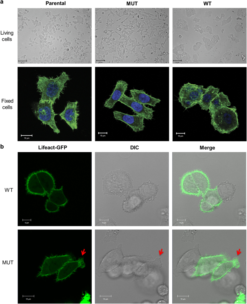
Figure 1.
Cell morphology of HCT116 cells is altered by the H1047R mutation in the p110α kinase domain of PI3K. (a) Cell morphology of HCT116 cells. Top panel: cell morphologies of live parental, WT and MUT HCT116 cells captured at a ×20 magnification. Bottom panel: confocal images parental, WT and MUT HCT116 cells captured at a ×63 magnification. Cells were fixed and stained for F-actin (green). Nuclei were stained with DAPI (blue). (b) Movement and morphology of live HCT116 WT (top) and MUT (bottom) cell at a ×40 magnification. Cells were transfected with Lifeact-GFP and cultured in a chambered cover glass dish for 24 h, and fluorescence and DIC images were acquired over 20 min.

