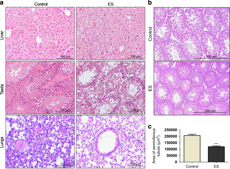
Figure 2.
Histopathological examination of organs from Endosulfan exposed mice and rat. (a) Histopathology of liver, testes and lungs of mice following ES treatment. Control in all panels indicates respective tissues from mice with no treatment and ES represents tissues from ES-treated mice (20×) (3 mg/kg, 1st day). (b) Histopathology of rat testes after ES treatment (10×) (3 mg/kg, 1st day). (c) Bar graph showing the relative difference in the diameter of seminiferous tubules of testes from control group and Endosulfan-treated rats (3 mg/kg). n=20 tubules in each group.

