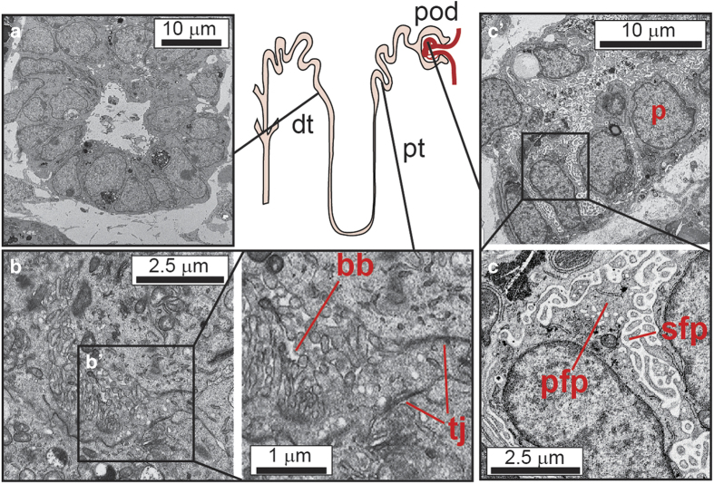
Figure 4.
Transmission electron microscopy showing the presence of specific nephron cell types within the kidney organoids generated from human-induced pluripotent stem cells. A diagram of the presumed location of specific cell types along the nephron is shown centrally. (a) Distal tubular epithelium showing clear lumen and small apical microvilli (b, bʹ) Proximal epithelial tubules showing evidence (bʹ) of brush border (bb) and cell–cell tight junctions (tj) (c, cʹ) Forming glomerulus with evidence of tightly interdigitated podocytes (cʹ) with primary (fpf) and secondary (sfp) foot processes.

