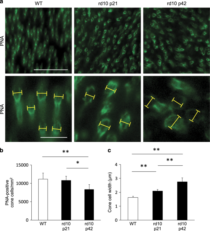
Figure 1.
Enlargement of the cone cell size in the mouse model of retinitis pigmentosa. (a) Whole-mount staining of the wild-type (WT; left panel) and rd10 mouse retinas at P21 (middle panel) and P42 (right panel) with PNA. The lower panels are higher magnification of peanut agglutinin lectin (PNA)-positive cone cells. Yellow line: the width of the inner segment of each cone cell. Scale bar, 50 μm (upper panel), 10 μm (lower panel). (b, c) Quantification of the density (b) and width (c) of PNA-positive cone cells in WT mice (n=6), rd10 mice at P21 (n=6), and rd10 mice at P42 (n=6). *P<0.05, **P<0.01.

