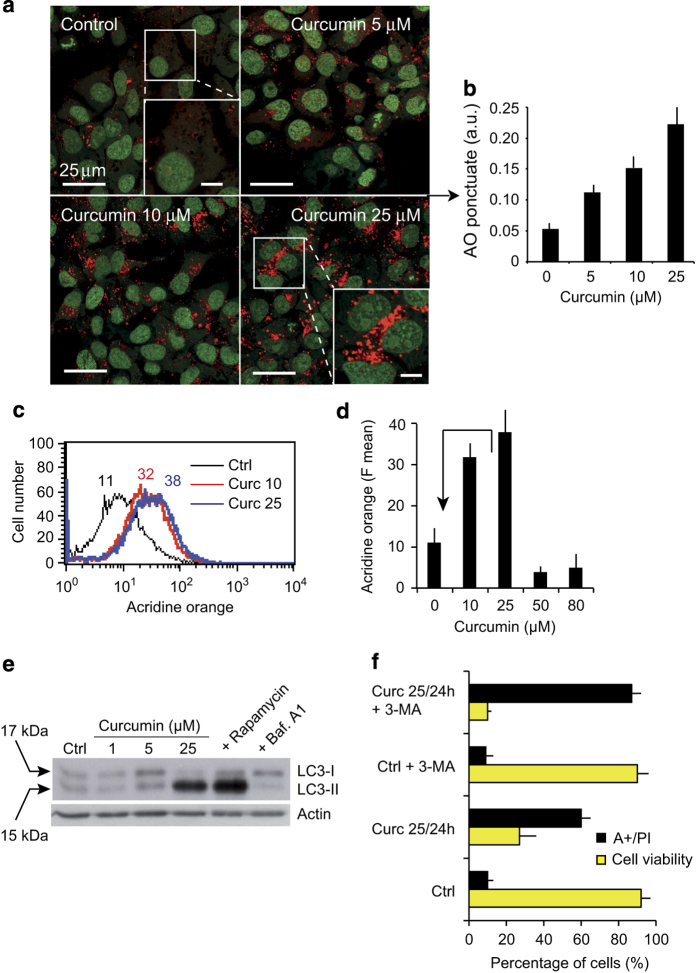
Figure 6.
Curcumin induce autophagy. (a) Confocal microscopy of AO-stained vesicles in cells treated with different concentrations of curcumin (5, 10 or 25 μM). In the section showing cells treated with 25 μM curcumin, the enlarged panel (lower right panel) reveals the massive accumulation of AO-positive vesicles. (b) Quantification of AO-punctuate staining (intracytoplasmic vesicles). The means (±S.D.) of seven independent experiments are shown **P<0.01. (c and d) Flow cytometric analysis of AO red fluorescence. In d, the mean fluorescence values±S.D. of seven independent experiments is shown. (e) Western blot analysis of the conversion of LC3-I to LC3-II in cells treated with different curcumin concentrations (1, 5 or 25 μM). Rapamycin was used to induce autophagy and Bafilomycin A1 to inhibit it. (f) Treatment with the autophagy inhibitor 3-MA promotes curcumin-induced apoptosis. Huh-7 cells were treated with curcumin (20 μM) and/or 3-MA (10 mM) for 24 h. Cell viability was determined by the PI staining assay, and apoptosis evaluated with Annexin V-FITC. Positively-stained cells were counted using a FACScalibur 4C. Data are expressed as the mean±S.D., n=6. **P<0.01 versus control; *P<0.05 versus curcumin-treated group.

