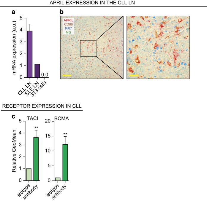
Figure 1.
APRIL is present in the CLL LN and CLL cells express APRIL receptors. (a) After total RNA lysis of paraffin-embedded LN material or control NIH-3T3 (3T3) cells, APRIL mRNA levels were determined by performing a qPCR on CLL LN material (N=3) and an SLE LN as positive and 3T3 cells as negative control. All qPCRs were performed in triplo. a.u. denotes arbitrary units. (b) Paraffin embedded LN slides from six CLL patients were immunohistochemically stained for APRIL, macrophage marker CD68, proliferation marker Ki67, and nuclear counterstain methyl green (MG). Data shown are representative of N=6. Scale bar represents 200 μm (left) or 50 μm (right). (c) CLL cells (N=6) isolated from PB were stained for APRIL receptors TACI and BCMA or with the relevant isotype controls and analyzed by flow cytometry. Bars show mean±S.E.M. **P<0.01 in a paired t-test.

