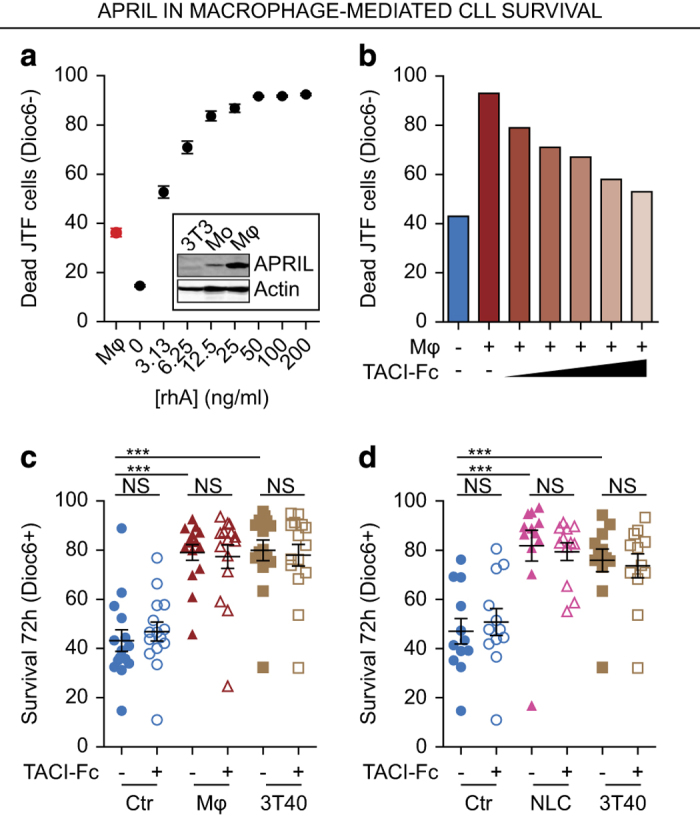
Figure 4.

APRIL is expressed by macrophages, but has no role in macrophage-mediated survival. (a) JTF reporter cells were stimulated for 24 h with different concentrations of rhA or with M1-differentiated macrophages. Consequently, cell viability was determined as in Figure 2d and the macrophage-induced cell death was plotted alongside of the rhA titration curve. All conditions were performed in triplo and mean±S.E.M. are shown. Inset: APRIL expression was determined in these macrophages (Mφ) by western blot and compared with monocytes (Mo) and untransduced 3T3 cells as negative control. (b) JTF reporter cells were stimulated with M1-differentiated macrophages as in Figure 4a in the presence of increasing concentration of the APRIL decoy receptor TACI-Fc (from 0.25 μg/ml to 2.5 μg/ml) or control IgG after which cell viability of the JTF cells was measured as in Figure 2d. (c) Confluent feeder layers of macrophages (Mφ) were generated as in Figure 4a and 3T40 feeder layers as in Figure 2e. These feeder layers or empty wells (Ctr) were then pre-incubated for 30 min with TACI-Fc to suppress APRIL signaling or control IgG after which CLL cells were added on these feeder layers and co-cultured for 72 h. Next, survival of the CLL cells was determined as in Figure 2e. Each point is one CLL sample (N=15) cultured in the indicated condition and mean±S.E.M. are indicated. ***P<0.001; NS, not significant, in an ANOVA test for repeated measures with Dunnett’s post hoc analysis. (d) Confluent feeder layers of NLCs were generated by differentiating monocytes for 10 days using CLL cells. After washing, their survival effect on CLL cells in the presence of absence of TACI-Fc was determined as in Figure 4c. Each point is one CLL sample (N=12) cultured in the indicated condition and mean±S.E.M. are indicated. ***P<0.001; NS, not significant, in an ANOVA test for repeated measures with Dunnett’s post hoc analysis.
