Figure 1.
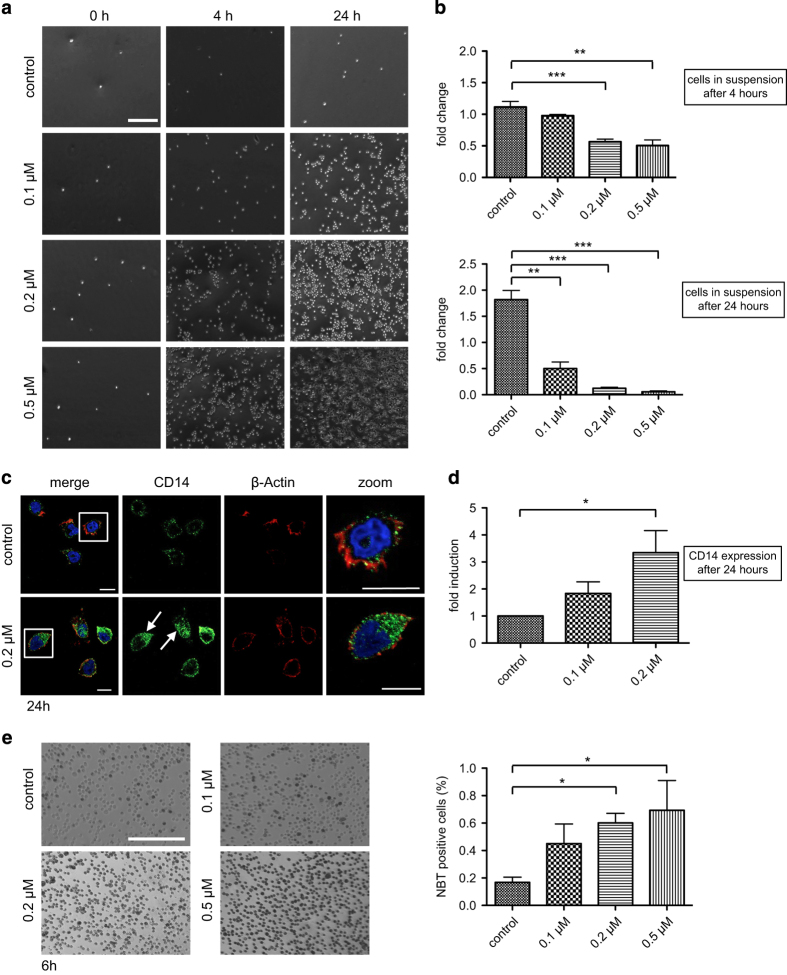
LecB induces differentiation of AML cells. (a) Light microscopy images of THP-1 cells treated with the indicated concentrations of LecB. After distinct time points, the cells remaining in suspension were removed and the adhered ones were illustrated. Scale bar, 250 μm. (b) The number of cells remaining in suspension upon LecB treatment was determined at indicated time points. Results are expressed as fold change of the cell amount present in the samples relative to the amount at time point 0. (c) Immunofluorescence images of THP-1 cells treated with 0.2 μM LecB for 24 h and subsequently stained with a CD14 specific antibody (green, arrows), Phalloidin-Atto-647 (red) and DAPI (blue). Scale bars, 10 μm. (d) Flow cytometry analysis of THP-1 cells treated with the indicated LecB concentrations for 24 h revealed a significant induction of CD14 expression. Results are expressed as fold change relative to the nontreated control sample. (e) Light microscopy images of THP-1 cells treated with the indicated concentrations of LecB and NBT substrate. After 6 h, the amount of cells bearing the insoluble blue intracellular accumulations were defined as NBT positive and quantified by ImageJ. The values are expressed relative to the total amount of cells. Scale bar, 200 μm. *P<0.05; **P<0.01; ***P<0.001.

