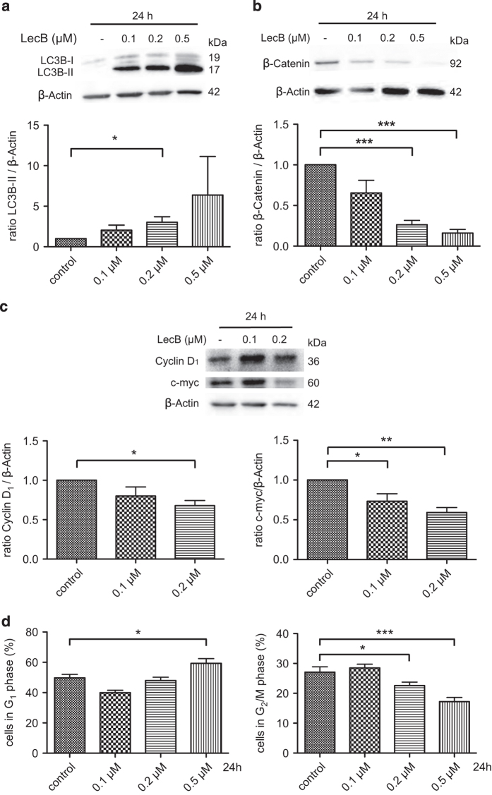
Figure 3.
LecB induces autophagy and reduction of β-Catenin in AML cells. (a) THP-1 cells were stimulated with the indicated concentrations of LecB for 24 h and the conversion of LC3B-I to the autophagosome marker LC3B-II (arrows) was analyzed in western blot experiments. Representative western blot images are illustrated. The quantification shows the fold increase of the LC3B-II normalized to β-Actin. (b) THP-1 cells were stimulated with LecB for 24 h and the β-Catenin protein level was determined in western blot experiments. Representative western blot images and their quantification are illustrated. The results illustrate the fold change of the β-Catenin level normalized to β-Actin. (c) Cells were stimulated with the indicated concentrations of LecB for 24 h and the expression of β-Catenin target genes Cyclin D1 and c-myc was analyzed in western blot experiments. Representative western blot images are illustrated. The quantification shows the fold change of the protein level normalized to β-Actin. (d) Treatment with LecB for 24 h affected the proliferation of THP-1 cells shown by cell cycle analysis with propidium iodide and measured by flow cytometry. *P<0.05; **P<0.01; ***P<0.001.

