Figure 6.
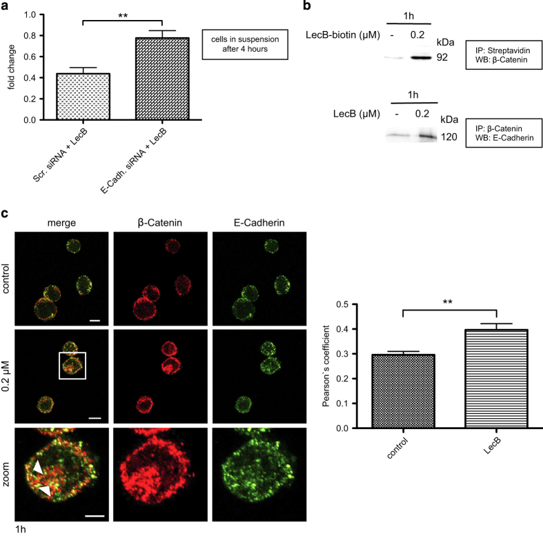
LecB strengthens the formation of E-Cadherin/β-Catenin complexes. (a) THP-1 cells were first transfected with the indicated siRNAs for 72 h and then treated with 0.2 μM LecB for 4 h. The number of cells remaining in suspension upon the treatment was determined. Results are expressed as fold change of the cell amount present in the samples relative to the amount at time point 0. (b) Western blot analysis of THP-1 cells stimulated with 0.2 μM LecB or biotinylated LecB for 1 h. Immunoprecipitations for streptavidin and β-Catenin were performed and the precipitated protein complexes were stained for β-Catenin and E-Cadherin, respectively. (c) Immunofluorescence images of THP-1 cells treated with 0.2 μM LecB for 1 h and subsequently stained with a β-Catenin- (green) and an E-Cadherin-specific antibody (red). Colocalizations are indicated with arrowheads. Scale bars, 10 μm (overview) and 5 μm (zoom). Colocalization was determined by Pearson’s coefficient, and the values represent the mean±S.E.M. of 10 analyzed cells per condition. **P<0.01.

