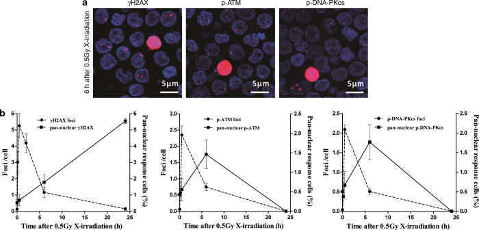
Figure 1.
Distinct kinetic patterns between the pan-nuclear responses and the repair foci of γH2AX, p-ATM and p-DNA-PKcs. (a) Representative images of X-irradiation-induced pan-nuclear signals and foci of γH2AX, p-ATM and p-DNA-PKcs in resting HPBLs at 6 h after 0.5 Gy X-ray exposure. ×60 magnification, ×1.0 zoom. (b) HPBLs were irradiated with 0.5 Gy X-ray, and the pan-nuclear signals and foci of γH2AX (left panel), p-ATM (middle panel) and p-DNA-PKcs (right panel) were examined at the indicated time points after irradiation and quantified by counting 1000–5000 HPBLs from each sample. γH2AX, p-ATM and p-DNA-PKcs were labeled in red, and the nuclei were stained in blue with DAPI. Data are presented as the means±S.D. of four to six donors.

