Figure 4.
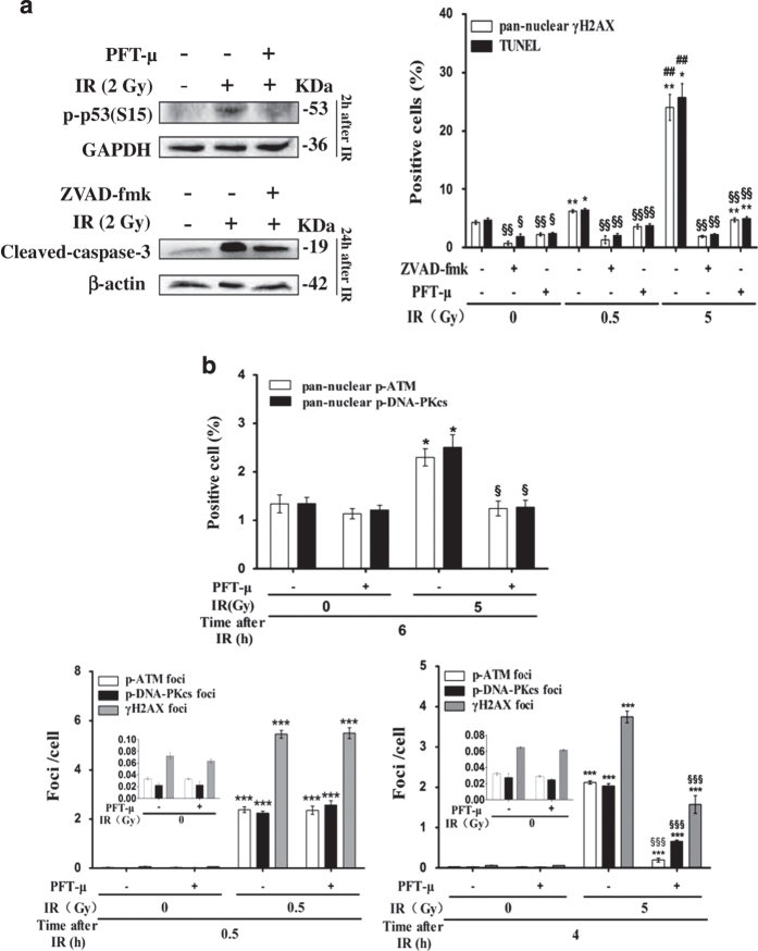
Inhibition of X-irradiation-induced pan-nuclear γH2AX response and apoptosis in resting HPBLs by PFT-μ and ZVAD-fmk. (a) HPBLs were treated with 20 μM PFT-μ for 5 h or 100 μM ZVAD-fmk for 1 h before X-irradiation (2 Gy). The levels of p-p53 expression (left upper panel) and cleaved caspase-3 expression (left bottom panel) in HPBLs at indicated time points post irradiation were examined by western blotting. Furthermore, pan-nuclear γH2AX and TUNEL-positive cells were counted at 24 h post irradiation. (b) HPBLs was treated with 20 μM PFT-μ for 5 h before X-irradiation. Pan-nuclear p-ATM- and p-DNA-PKcs-positive cells were counted at 6 h post irradiation (upper panel). γH2AX, p-ATM and p-DNA-PKcs foci were measured in HPBLs at 0.5 h (left bottom panel) and 4 h (right bottom panel) post irradiation. A total of 1000 HPBLs from each sample were examined for quantitation of the p-ATM, p-DNA-PKcs and γH2AX foci, and 2000–5000 HPBLs from each sample were examined to calculate the percentage of pan-nuclear γH2AX, p-ATM, p-DNA-PKcs and TUNEL-positive cells. The values shown in panel (a) and panel (b) represent the means±S.D. obtained from three to four donors. *P<0.05, **P<0.01 and ***P<0.001 compared with corresponding non-irradiated HPBLs with/without inhibitor treatment; ## P<0.01 compared with corresponding HPBLs exposed to 0.5 Gy X-ray irradiation with/without inhibitor treatment; § P<0.05, §§ P<0.01 and §§§ P<0.01 compared with corresponding HPBLs without inhibitor treatment.

