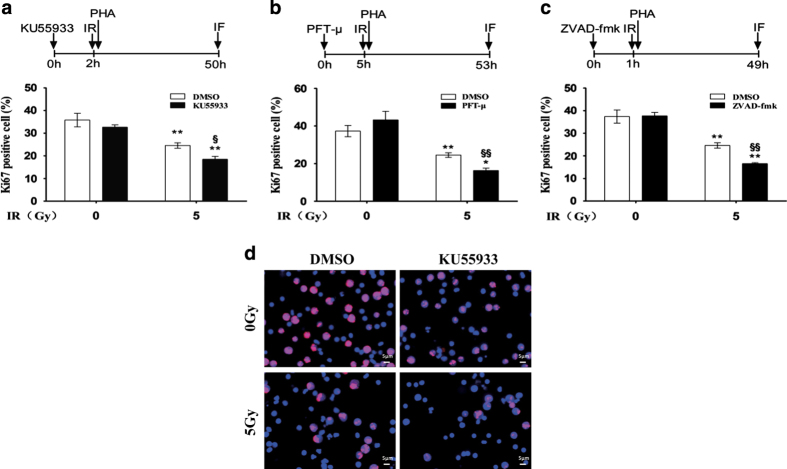
Figure 5.
KU55933, PFT-μ and ZVAD-fmk inhibited the proliferative response to mitogen in resting HPBLs exposed to X-ray irradiation. HPBLs were treated with 10 μM KU55933 for 2 h (a), 20 μM PFT-μ for 5 h (b) or 100 μM ZVAD-fmk for 1 h (c) followed by irradiation with 5 Gy X-ray, and were cultured with PHA and colcemid for 48 h. HPBLs were then collected and transformed HPBLs were detected using the Ki67 antibody. A total of 1000 HPBLs from each sample were examined to determine the proliferation ratio. Data are presented as the means±S.D. obtained from three donors. *P<0.05 and **P<0.01 compared with corresponding non-irradiated HPBLs with/without inhibitor treatment; § P<0.05 and §§ P<0.01 compared with corresponding HPBLs without inhibitor treatment. (d) Representative images of HPBLs labeled with Ki67. The cells were treated with KU55933 and irradiated with X-ray as described above. Ki67 was labeled in red and nuclei were stained in blue with DAPI. ×40 magnification, ×1.0 zoom.

