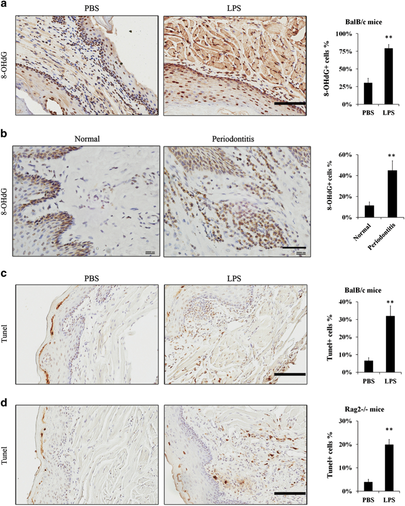
Figure 4.
Chronic periodontitis-induced apoptosis and DNA damage in gingiva. Approximately, 10 μg LPS in 10 μl PBS or 10 μl PBS vehicle were injected into mandibular buccal gingiva two times per week for 3 weeks to establish experimental periodontitis models or PBS vehicle models. (a) There were higher expressions of 8-OHdG in the nuclei in BalB/c mice periodontitis models compared to the PBS vehicle models (scale bar, 100 μm). (b) Paraffin-embedded human gingival samples were stained for 8-OHdG by IHC (other two healthy and four periodontitis specimens were shown in Supplementary Figure 1; scale bar, 50 μm). The percentage of positive cells was counted from four microscopic fields per sample. (c) TUNEL staining showed more apoptotic cells in BalB/c mice periodontitis model, especially in submucosal tissue (scale bar, 100 μm). (d) TUNEL staining in Rag2−/− mice periodontitis model (scale bar, 100 μm). The percentage of positive cells (a, c and d) was counted from three to four mice samples and four microscopic fields per sample (**P<0.01).

