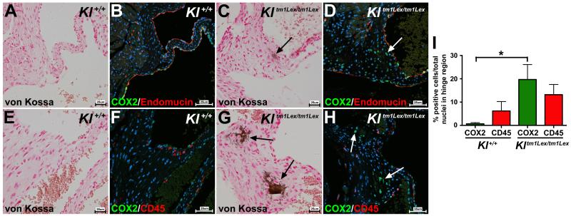
Figure 3. VICs expressing COX2 do not co-express endothelial or leukocyte/hematopoietic cell markers in klotho-deficient mice.
COX2 (green staining) is co-expressed with the endothelial marker endomucin (red staining) in endothelial cells in wild type animals (Kl+/+) (B). In klotho-deficient mice (Kltm1Lex/tm1Lex), in addition to endothelial cell expression, COX2 is evident in VICs in regions of calcification (COX2 expression indicated by white arrow in D), which are not positive for endomucin. Kl+/+ mice exhibit dispersed expression of the leukocyte/hematopoietic cell marker CD45 (red staining in F). In Kltm1Lex/tm1Lex mice, co-immunofluorescent staining of CD45 (red staining) and COX2 (green staining) shows that COX2-expressing cells do not express CD45 (COX2 staining is indicated by white arrows in H). Quantification of COX2-positive (green bars) and CD45-positive (red bars) VICs in the AoV hinge region demonstrates a significant increase in COX2 expression in Kltm1Lex/tm1Lex compared to wild type animals, whereas CD45 expression was unchanged (I). AoV calcification is detected by von Kossa staining in adjacent sections of the same specimens (brown staining indicated by black arrows in A, C, E, G; sections are counterstained with nuclear fast red). Nuclei are counterstained with ToPro3 (blue staining in B, D, F, H). A one-way ANOVA with a post-hoc multiple comparisons test was used to compare expression of COX2 and CD45 in wild type and klotho-deficient mice, a significant difference is indicated by an asterisk (p<0.05) and error bars represent the SD (I).

