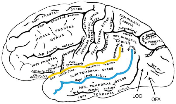
FIGURE 5.
Lateral view of the adult human brain (Greys Anatomy 726) with cortical areas labeled. Approximate locations of the lateral occipital complex (LOC) and occipital face area (OFA) are displayed. The fusiform face area (FFA) is located underneath the surface of the cortex, hence cannot be viewed here or investigated using fNIRS. The superior temporal sulcus is highlighted in red and the Sylvian fissure (or lateral sulcus) is highlighted in yellow.

