
Figure 4.
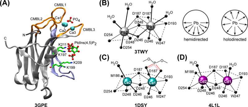
(A) The crystal structure of C2 domain from PKCα complexed to Ca2+ and PtdIns(4,5)P2.60 CMBL regions and the LRC lysine residues, K197, K199, K209, and K211, are highlighted with orange and blue, respectively. Adapted from61: coordination geometries of Pb2+ (B), Ca2+ (C), and Cd2+ (D) ions bound to the C2 domain from PKCα. The ligands are the sidechain oxygens of aspartates, with the exception of W247 and M186, where it is the carbonyl oxygen. The coordination sphere of Pb2 is hemi-directed: all eight ligands are located in one coordination hemisphere that is facing the viewer. The top axial ligands of Ca1 in 1DSY are the phosphoryl oxygens of the short-chain PtdSer analog.