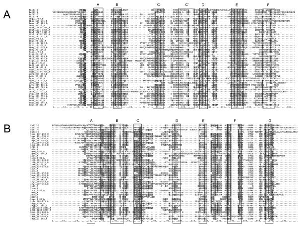
Figure 2.
Sequence alignment of the predicted domains in the REJ module to a set of sequences representative of the FNIII fold (A) and the Ig fold (B). The four putative domains from PC1 are labelled FNIII1-4. All other sequences are taken from structures available from the PDB. All of these are labelled by their PDB accession number, beginning and end of the domain in case of multidomain proteins and the molecule from which the sequence was taken. Expected β-strands for both folds are indicated by black boxes around the alignment which are labelled above. Sequence conservation is indicated by shading of residues (dark gray: hydrophobic; light gray: hydrophilic; Black with white character: proline).

