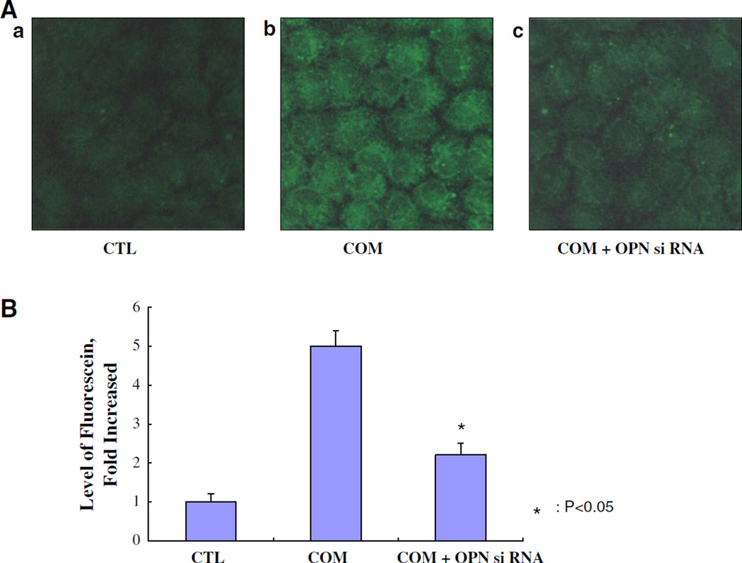
Fig. 1.
The transfection of OPN siRNA into NRK52E cells examined using siPORT™ NeoFX™ Transfection Agent. A a Control cells (CTL), b cells treated with calcium oxalate monohydrate (COM) crystals (66.7 µg/cm2) for 48 h, c cells with transfection of OPN siRNA treated with COM crystals. B Fluorescence of OPN levels was determined by confocal microscope images (LSM 5 Pascal, Carl Zeiss). Chemiluminescence intensities were calculated by Luzex® detection system (Nireco). The expression of OPN mRNA was significantly knocked down by OPN siRNA transfection (P < 0.05)

