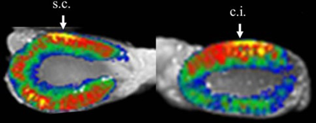
Fig. 2.
Fluorescence imaging; Alexa633-labeled atelocollagen (AteroGene™) was revealed between renal cortical injection (c.j.) and renal sub-capsular injection (s.c.) of atelocallagen. Atelocollagen injected in the renal cortex remained at the injection site after 24 h of the treatment, whereas atelocollagen injected at the sub-capsular site was visualized in the renal parenchyma far from the injection site. Image analysis was performed by macro-imaging station, BioView 1000 mounted onto a dark box and using 530/610 nm excitation/emission filters

