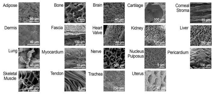
Figure 2.
Representative scanning electron microscopy (SEM) images for different decellularized tissues. These images demonstrate the vast differences in microenvironment architecture that are found in different tissues. These architectures make up the local niche that cells interact with and are therefore understood to play a significant role in shaping cell differentiation into corresponding cell lineages. SEM images reprinted with permission from [23, 31, 32, 36, 55–68].

