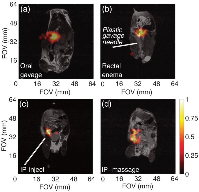
Fig. 7.
In vivo MRI for silicon particles. (a) Administration of of PEGylated silicon particles (in PBS) into a normal mouse via oral gavage, followed by a 5-min wait prior to imaging. (b) Administration of of PEGylated silicon particles (in 1 ml PBS) via injection through the rectum of a normal mouse using a soft plastic gavage needle (3.8 mm diameter), followed by a 5-min wait prior to imaging. (c) Administration of of PEGylated silicon particles (in PBS) via intraperitoneal injection into a normal mouse, followed by a 30-min wait prior to imaging; gravitational settling is noted at the injection site. (d) Administration of of ESTA-1 functionalized silicon particles (in PBS) into a SKOV3 mouse via intraperitoneal injection, followed by physical manipulation of the mouse’s abdomen to help distribute the particles, then a 10-min wait prior to imaging. imaging scans (color): 90 deg RARE imaging sequence, TR/TE: , FOV: , resolution: 2 mm; single scan acquisition (), processed with 35% threshold to filter background. Coregistered with imaging scans (grayscale): 90 deg RARE imaging sequence, coronal plane, TR/TE: , FOV: ; resolution: 0.25 mm; three averages ().

