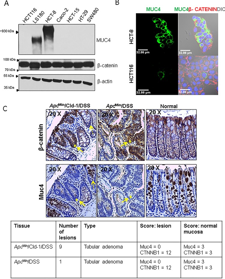
Figure 1. Increased nuclear β-catenin is associated with reduced MUC4 expression.
(A) A panel of CRC cell lines was profiled for the expression of MUC4 and β-catenin. β-actin was used as a loading control. (B) Confocal microscopy with MUC4 (green) antibody shows that HCT-8 cells express MUC4 abundantly while HCT116 cells show very low MUC4 expression. β-catenin (red) is present in both cell lines.(C) Immunohistochemical staining for mouse Muc4 and β-catenin in colon sections from ApcMin/Cldn-1 mice treated with DSS (left panel), ApcMin mice treated with DSS (middle) and ApcMin given water (right panel). Staining for β-catenin (upper panel) and Muc4 (lower panel) showed intense cytosolic/nuclear staining for β-catenin and depletion of Muc4 in lesions (solid arrow), while surrounding normal areas showed reduced β-catenin and intense goblet cell staining for Muc4 (dotted arrow). Table shows type, number of lesions in in mice either treated with DSS alone or ApcMin mice treated with DSS.

