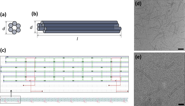
Figure 1.
Six-helix bundle. The (a) front and (b) side view of the six-helix bundle are schematically depicted (diameter: d = 6 nm, length: l = 414 nm). (c) caDNAno design representation with the template strand in blue, and main and capture staples in green and red, respectively. The capturing strands are 126 bases apart from each other on both helices. (d,e) TEM images of the resulting tubular DNA structures. The scale bar is (d) 200 nm and (e) 100 nm.

