
Figure 5.
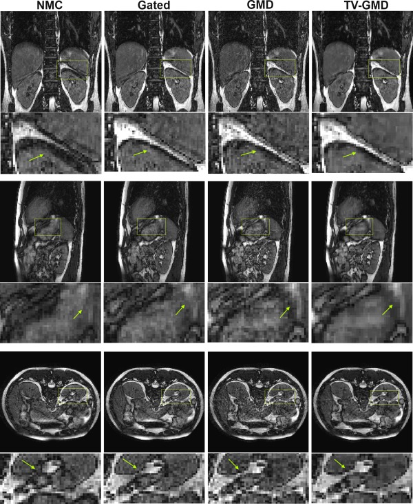
Coronal (top), sagittal (middle) and axial (bottom) slices for volunteer 1 with maximum respiratory amplitude of 14.8 mm (including zoom‐in images, arrows point out some main differences). NMC (non‐motion corrected): 2× undersampled at 100% gating efficiency. Several structures in the image are corrupted by motion. Gated: 2× undersampled at 60% gating efficiency. Most structures are sharper than the NMC. GMD: 3.5× undersampled at 80% gating efficiency. The GMD is sharper than the NMC, but presents remaining undersampling artifacts. TV‐GMD: 3.5× undersampled at 80% gating efficiency. The total variation regularization improves undersampled reconstruction at the expense of minor blurring.