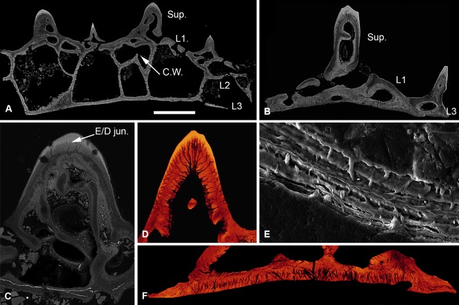
Figure 2.

Histology of Loricopteraspis serrata. NRM‐PAL C.5939, SEM BSE section through the cephalothoracic shield (A); NRM‐PAL C.5940, SRXTM section through a scale unit (B); NRM‐PAL C.5941, SEM BSE section through a tubercle, showing the enemeloid capping layer (C); NRM‐PAL C.5940, SRXTM volume rendered virtual thin section of a tubercle, showing the arrangement of dentine canaliculi radiating from a pulp canal (D); NRM‐PAL C.5942, etched SEM section through a wall of L2, showing homogenous core and lamellar margins pervaded by an orthogonal fabric of thread‐like spaces (E); NRM‐PAL C.5940, SRXTM volume rendered virtual thin section of L3, showing Sharpey's fibres trending in two principal orientations (F). Sup., superficial layer; L1, layer 1; L2, layer 2; L3, layer 3; C.W., incomplete cross wall; E/D jun., Enameloid/dentine junction. Scale bar equals 606 μm in (A), 227 μm in (B), 179 μm in (C), 55 μm in (D), 30 μm in (E) and 56 μm in (F).
