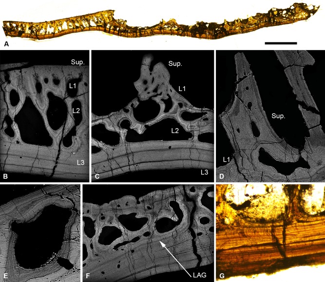
Figure 5.

Light microscopy thin section (A, G) and SEM BSE polished sections (B–F) through the cephalothoracic shield of Phialaspis symondsi, NHM P.73619. Section from the centre to the margin of the shield, showing the transition from the central ‘plate’ to the peripheral tuberculated region (A); histological structure of the central ‘plate’ (B) and peripheral region (C); detail of the superficial layer/L1 of the peripheral region. L1 centripetal lamellae are truncated, suggesting the vascular canals were remodelled via resorption (D); osteon lamellae of L2 truncated by vascular space, indicating resorption (E); section through the peripheral region, showing line of arrested growth demarking the previous margin of the shield (F), L3, showing alternating light and dark bands (G). Scale bar equals 3.3 mm in (A), 567 μm in (B), 523 μm in (C), 263 μm in (D) 113 μm in (E), 403 μm in (F) and 279 μm in (G).
