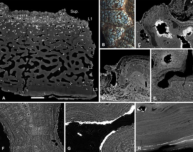
Figure 10.

Histology of Psammosteus megalopteryx, NHM P.73624. SEM BSE sections (A, C–H) and LM image (B). Structure of the cephalothoracic dermal skeleton (A), detail of surface ornament consisting of concentric tubercle ‘islands’ separated by grooves (B); section through a tubercle, showing centripetal dentine lamellae pervaded by polarised canaliculi (C); superposition of a second generation of tubercles. The tubercle on the right is completely in filled with secondary dentine (D); section through L1 showing truncation of lamellae by the vasculature, interpreted as evidence of resorption (E); section through a radial wall of L2, showing centripetal apposition of lamellae about a homogenous core. A fine fabric of orthogonal thread‐like spaces passes through and warps the lamellae (F); lamellae truncated by the vascular space, suggesting resorption of L2 (G); detail of L3 (H). Scale bar equals 785 μm in (A), 1.9 mm in (B); 99 μm in (C), 84 μm in (D); 121 μm in (E), 43 μm in (F); 41 μm in (G); 157 μm in (H).
