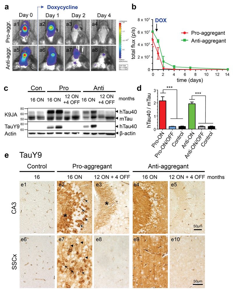
Fig. 2. Characterization of human full-length Tau (hTau40) expression in pro- and anti-aggregant transgenic mice.
(a) In vivo bioluminescence imaging (BLI) of luciferase activity. After addition of doxycycline (DOX, 200mg/kg) in food pellets, the transgene expression is down regulated within 4 days in pro-aggregant (a1-a4) and anti-aggregant mice (a5-a8). (b) Quantification of luciferase activity by in vivo BLI displayed in photons/s (p/s) after addition of DOX (mean ± SEM, n = 5 mice per group). (c) Representative expression of hTau40 (Mr ∼67kDa) and endogenous mouse Tau (mTau, Mr ∼45-55kDa) in pro- and anti-aggregant mice. Four months after switch-OFF, a loss of hTau40 is detected by the pan-Tau antibody K9JA and the human Tau specific antibody TauY9, β-actin serves as loading control. (d) Quantification of (c). Bars indicate the protein ratio of hTau40:mTau (mean + SEM). Pro-ON (n = 12) and anti-ON mice (n = 12) show a ∼2-fold overexpression of hTau40 over mTau as compared to pro-ON/OFF (n = 10), anti-ON/OFF (n = 10) and controls (n = 8). ***p<0.0001, one-way ANOVA with post-hoc Bonferroni´s multiple comparison test. (e) Distribution of hTau40 (antibody TauY9) in neurons of the CA3 region (e1-e5) of the hippocampus and somatosensory cortex (SSCx, e6-e10) in control, pro-ON, anti-ON, pro-ON/OFF and anti-ON/OFF mice. Note a mosaic like expression and localization of hTau40 to the cell somata (arrows), stratum lucidum (star) and apical dendrites (arrowheads) in pro-ON (e2, e7), whereas anti-ON shows a more diffuse distribution of hTau40 (e4, e9). After switch-OFF, staining intensities in pro-ON/OFF (e3, e8) and anti-ON/OFF (e5, e10) are clearly diminished. Scale bar 50µm.

