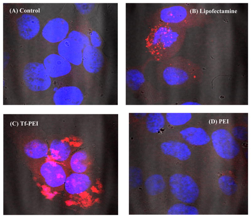
Figure 9.

Confocal microscopy images of cellular interaction of different complexes with the HCC1954 cell line after 4 h incubation with siRNA polyplexes. HCC1954 cells were seeded with 25,000 cells per chamber and transfected with 25 pmol of siRNA (TYE 563 labeled, indicated by red color) for 4 h. Cells were fixed and the nuclei stained with DAPI (indicated by blue color). Representative pictures are shown (100× magnification): (A) blank; siRNA polyplexes with (B) Lipofectamine; (C) Tf–PEI (N/P 7); and (D) PEI (N/P 7).
