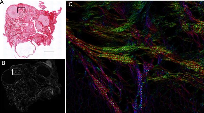
Figure 3. Picrosirius Red fibers in the aged ovary are birefringent.
A PSR-stained ovarian tissue section from a 22-month old CD1 mouse visualized by both (A) brightfield microscopy and (B, C) circularly polarized light microscopy (Abrio LCPolScope). The boxed region in (A, B) is magnified in (C) where the false color image with retardance vectors (green lines) reflects the orientation of the co-aligned fibers. The scale bar is 0.4 mm.

