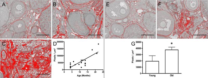
Figure 5. Fibrosis significantly increases in the ovarian stroma with advanced reproductive age in both CD1 and CB6F1 mice.
Representative processed color threshold images of PSR-stained ovarian tissue sections used to quantify fibrosis from CD1 mice that were (A) 4-months old, (B) 13.5-months old, and (C) 22-months old. (D) Graph showing the relationship between CD1 mouse age (months) and the average area of PSR-positive staining per ovarian section (pixels/μm2). A significant linear relationship exists between these variables (Pearson's correlation, P < 0.0001 and R2 = 0.6413). Representative processed color threshold images of PSR-stained ovarian tissue sections used to quantify fibrosis from CB6F1 mice that were (E) 6-12 weeks old (young) and (F) 14-17 months old (old). (G) Graph comparing the average area of PSR-positive staining per ovarian section (pixels/μm2) between reproductively young and old CB6F1 mice. The asterisk indicates a significant difference (P = 0.03). In all images, the red corresponds to PSR-positive staining, and scale bars are 200 μm.

