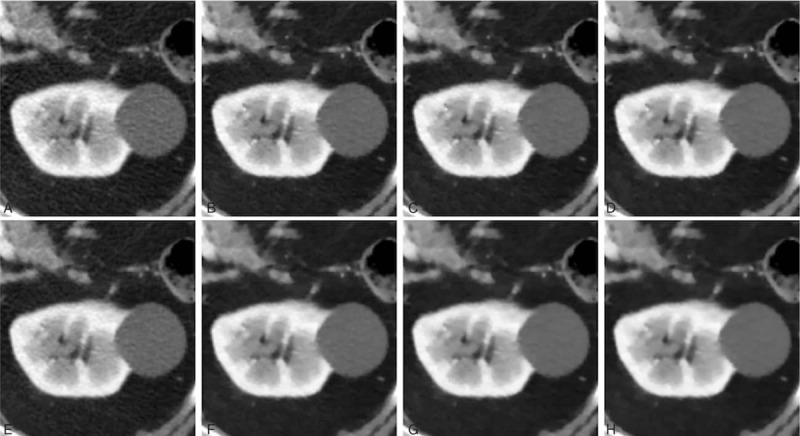
Figure 3.

Comparison of abdominal computed tomography image quality focusing on a renal cyst in an 83-year-old male patient. (A) FBP; (B) IMR-R-L1; (C) IMR-R-L2; (D) IMR-R-L3; (E) iDose4; (F) IMR-ST-L1; (G) IMR-ST-L2; and (H) IMR-ST-L3. The 3.4-cm renal cyst appeared inhomogeneous in the FBP (A) and iDose4 (E) reconstruction algorithms. Noise was reduced after applying IMR (B–H), yielding smoother images. FBP = filtered back projection, IMR = iterative model reconstruction, L = level, R = routine, ST = soft tissue.
