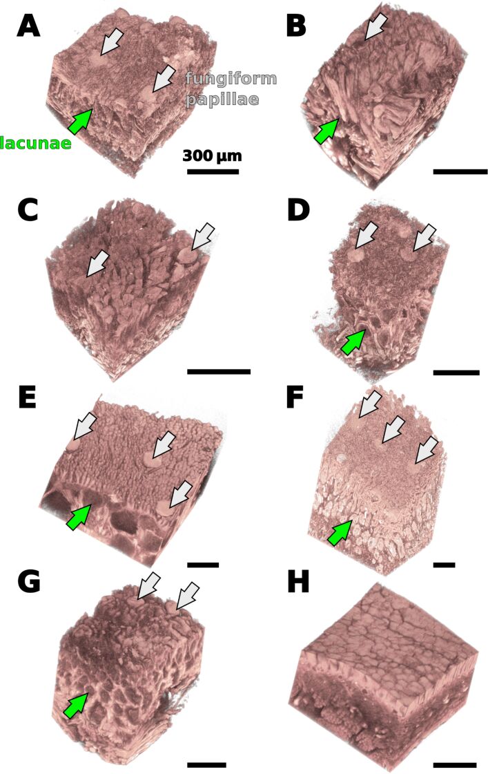
Figure 3.
Micro-CT images of tissue fragments that were derived from the surfaces of frog tongues. A – Bombina variegata, B – Discoglossus pictus, C – Ceratophrys ornata, D – Litoria caerulea, E – Megophrys nasuta, F – Rana (Lithobates) pipiens, G – Bufo bufo, H – Oophaga histrionica. Except for C. ornata (C) and O. histrionica (H), the fungiform papillae (grey arrows) can easily be identified in the micro-CT data. Underneath the papillary surface structures lies a layer with lacunar structures that appear hollow in the micro-CT scan (green arrows). Both size and shape of these lacunae strongly vary among different species.

