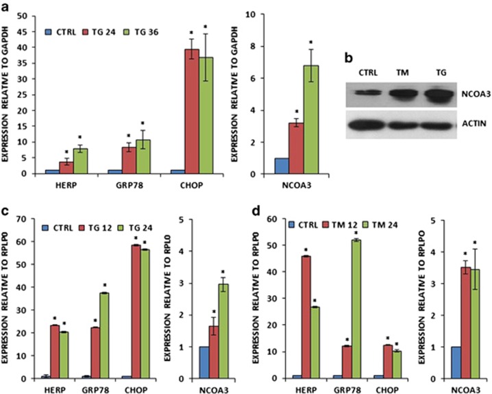
Figure 1.
Increased expression of NCOA3 by UPR in human breast cancer cells. (a) MCF7 cells were either untreated (CTRL) or treated with (1.0 μm) TG for indicated time points. The expression level of UPR-responsive genes (GRP78, HERP and CHOP) and NCOA3 was quantified by real-time RT–PCR, normalizing against GAPDH. Error bars represent mean±s.d. from three independent experiments performed in triplicate. (b) MCF7 cells were either untreated (CTRL) or treated with (1.0 μm) TG and (1.0 μg/ml) TM for 24 h. Western blotting of total protein was performed using antibodies against NCOA3 and β-actin. (c, d) T47D cells were either untreated (CTRL) or treated with (1.0 μm) TG (c) and (1.0 μg/ml) TM (d) for indicated time points. The expression level of UPR-responsive genes (GRP78, HERP and CHOP) and NCOA3 was quantified by real-time RT–PCR, normalizing against RPLP0. Error bars represent mean±s.d. from three independent experiments performed in triplicate. *P<0.05, two-tailed unpaired t-test compared with untreated cells.

