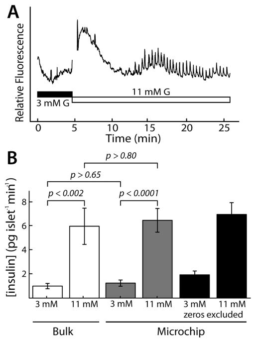
Figure 3.
Validation of passive microfluidic method for islet secretion sampling. (A) Synchronized calcium oscillations were observed in islets trapped in the microfluidic system after exposure to stimulatory glucose (11mMG), confirming that intercellular communication was intact. (B) Microfluidic secretion sampling was validated. Average insulin secretion from islets in the device (gray bars; 62 islets, 5 mice) was statistically equal to that measured by multi-islet, static methods (“Bulk” ; white bars; 200 islets, 5 mice). Black bars show microchip analysis when excluding undetectable secretion from 11 islets (less comparable to bulk methods). Error bars represent standard error of means of ELISA measurements for each islet or tube.

