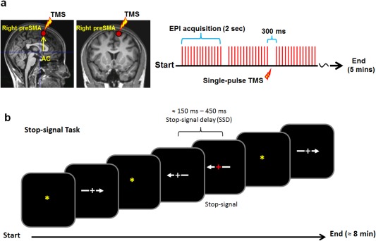
Figure 1.

(a) Localization of the TMS target (i.e., the right preSMA) and a schematic illustration of the timing of the single‐pulse TMS relative to the EPI acquisition sequence during the scans. Single‐pulse TMS was delivered 150 ms after the onset of the silence period (300 ms). The rfMRI scans were acquired using a gradient echo‐planar imaging (EPI) sequence with a TR of 2000 ms and a scanner silence period of 300 ms at the end of each TR. (b) Stop‐signal task applied in the study. The stop‐signal delay (SSD) was dynamically controlled such that it increased 50 ms for every successful stopping (stop‐inhibit) response and decreased 50 ms for each failed‐to‐stop (stop‐respond) response.
