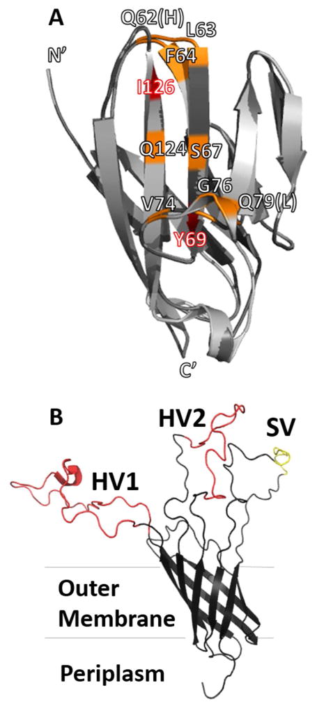
Figure 1.
(A) Structure of NCCM1 (PDB ID 2GK242, light gray) and homology model of NCCM3 (generated with SWISS-MODEL60–63, dark gray). Residues that bind to all OpaCEA proteins are colored red, while residues that only bind specific Opa variants are colored orange. All amino acids on the binding face of NCCM1 are conserved in NCCM3, except Gln62 (His in NCCM3), and Gln79 (Leu in NCCM3). (B) Structure of Opa60 (PDB ID 2MLH)30. The conserved regions of the protein are shown in black, hypervariable regions (HV1 and HV2) are colored red, and semi-variable region (SV) is colored yellow.

