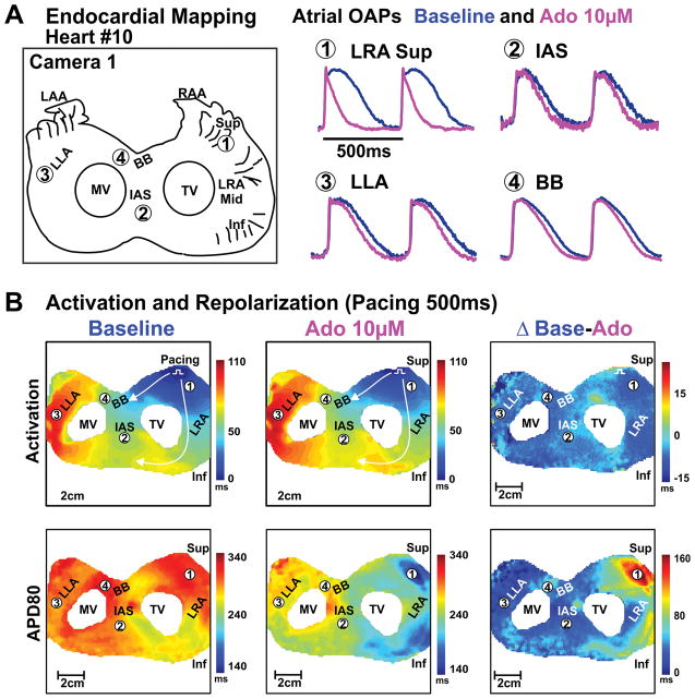
Figure 2. Endocardial optical mapping of bi-atrial preparation from failing human hearts.
A, Schematic of the optical field of view and optical action potentials from different atrial regions at baseline and 10μM adenosine perfusion of human Heart #10(522421). B, Activation and APD80 maps of the atria at baseline and 10μM adenosine perfusion; right panel shows the differences of activation and repolarization time between baseline and adenosine perfusion. Abbreviations as in Figure 1. Inf – inferior; LLA – lateral left atria; Mid – middle; MV – mitral valve; OAP– optical action potential; Sup – superior; TV – tricuspid valve.

