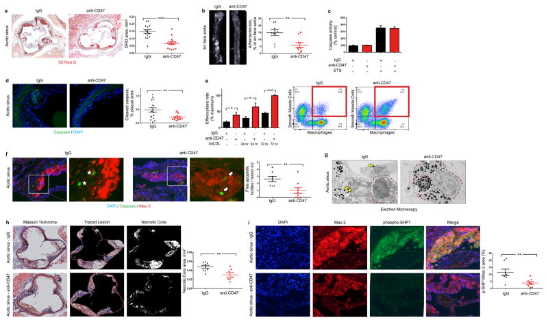
Figure 2. Inhibition of CD47 stimulates efferocytosis and prevents atherosclerosis.
(a). Compared to mice treated with a control Ab (IgG, n=16), mice treated with an inhibitory anti-CD47 Ab (n=15) develop significantly smaller atherosclerotic plaques, as measured by Oil-Red-O (ORO) content in the aortic sinus. (b). Total aortic atherosclerosis content is also reduced. Inhibition of CD47 signaling does not alter the rate of programmed cell death in vitro (c) but does reduce the accumulation of apoptotic bodies in vivo (d). (e). Anti-CD47 Ab promotes efferocytosis of vascular cells at baseline and after exposure to pro-atherosclerotic lipids. Representative FACS phagocytosis plots for lipid loaded (72hr) SMCs shown at right (all assays repeated in triplicate). (f). In vivo, anti-CD47 Ab reduces the number of ‘free’ apoptotic bodies not associated with phagocytic macrophages, potentially indicative of increased efferocytosis (stars indicate ‘free’ apoptotic bodies, arrows indicate ‘not-free’ apoptotic bodies). (g). Electron microscopy confirms that mice treated with anti-CD47 Ab display features of enhanced intraplaque efferocytosis, including an increased prevalence of macrophages which had ingested multiple apoptotic bodies (white arrows) and a reduced burden of ‘free’ apoptotic bodies (yellow arrows). (h). Mice treated with anti-CD47 Ab develop smaller necrotic cores than mice treated with IgG. (i). Anti-CD47 Ab inhibits phosphorylation of lesional SHP1, a key anti-phagocytic effector molecule known to signal downstream of CD47. STS = staurosporine. Comparisons made by two-tailed t tests. *** = P < 0.001, ** = P < 0.01, * = P < 0.05. Error bars represent the SEM.

