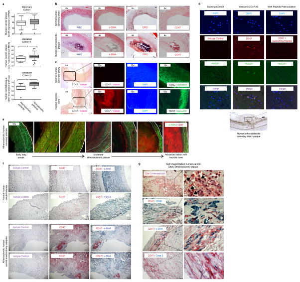
Extended Data Figure 1. CD47 expression correlates with risk for clinical cardiovascular events and is progressively upregulated in the necrotic core of human blood vessels during atherogenesis.
(a). cDNA microarray expression profiling in the BiKE carotid endarterectomy biobank reveals that the relative expression of CD47 is increased in vascular homogenates taken from subjects with symptomatic disease (stroke or TIA, n=85) compared to those with stable, asymptomatic lesions (n=40). Similar findings were observed in the non-overlapping discovery and validation cohorts from BiKE (n=55), and a second validation cohort from the Helsinki Carotid Endarterectomy Study (HeCES, n=21). Data presented as Tukey boxplots. (b). Immunohistochemical staining reveals that CD47 co-localizes with lipidated plaque within human coronary lesions, as measured by Oil-Red-O (ORO) staining. (c). Immunofluorescent staining of coronary samples confirms that CD47 is upregulated within the necrotic core. (d). High magnification (40x) imaging of atherosclerotic coronary plaque confirms that CD47 expression is present on the surface of nucleated cells undergoing cell death, as indicated by HMGB1 staining. Specificity of the anti-CD47 Ab is confirmed in assays where the signal was quenched by preincubating the sections with recombinant CD47 peptide prior to primary antibody exposure. (e). Additional representative coronary artery segments spanning the spectrum of progressive coronary artery disease (non-atherosclerotic coronary, early ‘fatty streak’, inwardly remodeled plaque, and advanced ulcerated lesion with necrotic core) confirm that CD47 is progressively upregulated during the development of coronary artery disease. The tunica media is indicated by dotted lines. (f). Additional staining in human carotid artery sections confirms that CD47 expression is upregulated in atherosclerosis relative to healthy tissue, and appears most pronounced within the necrotic core. (g). High magnification (100x) imaging confirms that the CD47 expression is specific to lesional cells, including SMCs (αSMA), macrophages (CD68) and cells undergoing programmed cell death (Casp3). Comparisons made by two-tailed t tests. ** = P < 0.01, * = P < 0.05.

