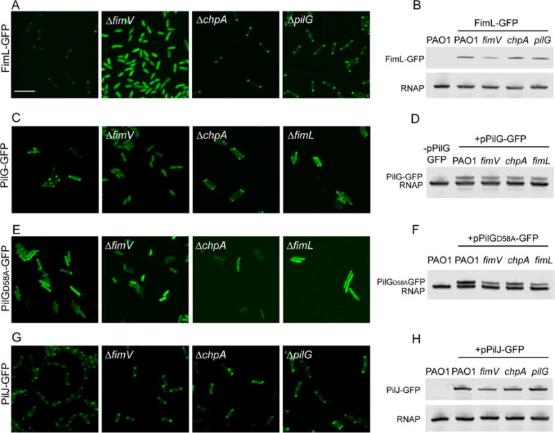
Fig. 5.

Subcellular localization of FimL-GFP, PilJ-GFP and PilG-GFP. The indicated PAO1 strains expressing (A) FimL-GFP from the chromosome, (C) plasmid-encoded PilG-GFP (pPilG-GFP), (E) plasmid-encoded PilGD58A-GFP (pPilGD58A-GFP) or (G) plasmid-encoded PilJ-GFP (pPilJ-GFP) in wild-type PAO1, PAO1ΔfimV, PAO1ΔchpA and PAO1ΔfimL mutants were grown on agar pads with 0.02% arabinose induction for 4 h and then examined by confocal microscopy. Scale bar represents 5 μm. (B,D,F,H) Immunoblot of lysates prepared from the indicated strains probed with the anti-GFP (top band) or anti-RNAP, a loading control (bottom band).
